Western Blotting
Discover our comprehensive range of western blotting solutions for optimal transfers of proteins and nucleic acids from electrophoresis gels onto carrier membranes.


Western Blotting
Western blotting (or the western blot) is a commonly used technique for analyzing proteins. This method makes it possible to identify specific proteins from a complex of proteins by transferring the target proteins to a carrier membrane (blotting). Proteins are first separated into protein bands via gel electrophoresis and transferred to a carrier matrix with an electric field, where they bind in different ways depending on their properties and where they can be detected using various methods.
Western blotting is a core method in pharmaceutical, biochemical and medical research; and it plays an essential role in diagnosing viral infections. As one of the most widely used methods for protein analysis, the method is prized for its simple, cost-effective and highly specific identification of proteins.
The Western Blot - Origin and History of the Term
The term "western blotting" (where "blotting" means "transfer") is a play on the name of the Southern blot: The Southern blot is a similar method for detecting specific DNA sequences, pioneered by Ed Southern.
In addition to the western blot, the "northern blot" has become an established term, referring to the corresponding separation of RNA fragments. The following overview shows blotting methods with similar names, each of which describes a different analytic method in molecular biology:
- Southern blot: A method established by Ed Southern for separating DNA fragments, with subsequent hybridization.
- Northern blot: The Southern blot's counterpart for splitting RNA fragments.
- Western blot: The transfer of proteins to a carrier membrane as a method for analyzing proteins.
- Far-western blot: A method for investigating protein-protein interactions.
- Southwestern blot: A method for detecting DNA-protein reactions.
- Northwestern blot: A method for detecting RNA-protein reactions.
The term "western blot" was coined in 1981 by W. Neal Burnette, although the method had already been discovered independently two years earlier by scientists at Stanford University and the Friedrich Miescher Institute in Basel.
Today, the method is one of the most widely used routine techniques for protein analysis and is used for both qualitative and semi-quantitative analysis.
The Blotting Principle and Protein Detection
The principle of western blotting relies on first using gel electrophoresis to separate proteins into protein bands based on properties like charge and size, then applying the protein mixture to be analyzed to a carrier matrix. This procedure makes the proteins accessible for detection using antibodies. The three main steps – gel electrophoresis, blotting and detection – are explained in detail below.
Step 1: Gel electrophoresis
Gel electrophoresis acts to separate the protein mixture depending on the properties of the proteins in the mixture. Various methods are used, which differ in terms of the nature of the gel and interaction with the gel. A widely used method is the SDS-PAGE method, in which a discontinuous polyacrylamide-based gel is used. This method unfolds and negatively charges the proteins and thus enables the separation of the proteins according to their chain length, which in turn is proportional to their molecular mass.
The polyacrylamide gel is placed in an ionic buffer solution and is subjected to a vertical electric field. The proteins migrate through the gel in the direction of the positively charged anode, ending up in different positions based on their molecular size. Smaller proteins migrate faster through the gel, resulting in separation according to protein mass.

Step 2: Blotting
During the actual blotting process, the sorted proteins are transferred to a membrane, which can be a membrane made of polyvinylidene difluoride (PVDF), nitrocellulose (NC), nylon or glass fiber, for example. PVDF and NC carriers are particularly common in protein analysis. NC membranes are characterized by a high protein affinity, but suffer from the disadvantage of quickly becoming brittle. In contrast to NC, PVDF has the advantage that the blot can be used for re-probing.
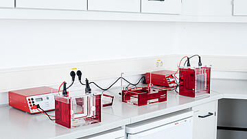
The transfer (blotting) of the proteins from the gel to the carrier matrix often takes place by means of electroblotting. Here, an electric current is applied to draw the negatively charged proteins into the membrane. In the experimental setup, the membrane is placed on the gel toward the anode side, with buffers on both sides in the form of wetted filter papers. The structure and size of the transfer buffer vary, but the following blot systems are widely used in practice:
- Tank blot systems (e.g. Biometra Tankblot Ecoline)
- Semi-dry blotting systems (e.g. Biometra Fastblot)
- Dry blotting systems
When the proteins are transferred, they retain their electrophoretic separation in the gel and are now present on the carrier matrix in their separated form. This makes them accessible for detection methods.
")
Figure 1: Diagram of a western blot transfer (Source: Bensaccount at English Wikipedia)
Step 3: Detection
After this, antibodies that bind to a specific protein are used to detect the proteins on the carrier matrix. This is how it is possible to identify, for example, viral proteins and thus to detect infections.
Before the proteins can be detected, interactions between the membrane and the detection antibody must be prevented. For this purpose, the membrane is placed in a diluted protein solution, for example 3–5% bovine serum albumin (BSA) or fat-free powdered milk. The solution binds the membrane at the sites where the target proteins have not attached – as a result, the antibodies can only bind to the specific target protein, which leads to a better analytic result.
The target protein can then be identified with a color reaction. This entails applying two different antibodies to the membrane. First, the primary antibody binds to the target protein. Then, non-target antibodies are rinsed from the membrane before the secondary antibody is applied. The secondary antibody docks at the so-called Fc fragment of the primary antibody and acts as a catalyst for the dye reaction when the right substrate is added. And finally, observing the color reaction allows detection of a specific antibody.
Areas of Application for Western Blotting
Western blotting has established itself in pharmaceutical and medical analysis as a reliable method for identifying specific proteins in complex mixtures of proteins. The method allows not only the qualitative detection of a protein, but also enables semi-quantitative estimation based on the size and color intensity of the protein band.
In protein biochemistry, Western blotting is used for the identification of proteins or protein modifications; for example, it is suitable for detecting post-translational modifications of proteins. In the medical field, western blotting has proven itself as a diagnostic method for various infectious diseases. For example, the method allows for detection of antibodies in blood serum, such as in HIV tests. In cancer research, the western blot helps us evaluate drugs by quantifying ERK proteins and thereby assessing their effect on the growth of tumor cells.
Western blotting is often used in combination with other methods for the detection of proteins. For example, it is used as a test to confirm a positive ELISA screening test in HIV diagnostics.
The newsletter of Analytik Jena frequently keeps you posted about:
- News
- Trends and developments
- Events