

Polyacrylamide Gel Electrophoresis
Discover our comprehensive range of polyacrylamid gel electrophoresis (PAGE) and blotting solutions for optimal proteins and nucleic acid analysis.


Polyacrylamide Gel Electrophoresis
Polyacrylamide gel electrophoresis (PAGE) is one of the well-established standard methods in biochemical and molecular biology analysis for the precise separation of proteins and nucleic acids based on their molecular size and charge. The technique is based on the movement of electrically charged molecules through a porous gel under the influence of an electric field. Smaller molecules migrate more rapidly through the pore structure of the polyacrylamide gel than larger ones, resulting in a reliable and reproducible separation.
Thanks to its high resolution and robustness, polyacrylamide gel electrophoresis is particularly well suited for demanding analytical tasks in everyday laboratory work. Especially when dealing with complex samples composed of a wide variety of different molecules, PAGE enables clear differentiation and identification of individual components. Due to its broad range of applications, the method has become indispensable in both basic research and in clinical diagnostics, biotechnology, and pharmaceutical research.
Overall, the continuous advancement of these instruments and techniques plays a key role in making laboratory operations more efficient and improving the quality of analytical results. The following sections provide a closer look at the preparation and properties of polyacrylamide gels, the practical implementation of the method, and its wide range of applications.
Preparation and Properties of Polyacrylamide Gels
Polyacrylamide gels are formed through a controlled polymerization reaction in which the two monomers acrylamide and N,N'-methylenebisacrylamide react with each other. During this process, the bisacrylamide cross-links the linear polyacrylamide chains into a three-dimensional network, with the pore size determined by the ratio of the two monomers. Polymerization is initiated by free-radical starters such as ammonium persulfate (APS) and a catalyst, typically tetramethylethylenediamine (TEMED).
A key advantage of polyacrylamide gels over other gel materials, such as agarose, is their exceptionally high mechanical stability and chemical neutrality. This makes them particularly suitable for the separation of smaller molecules that require high resolution and sharp band separation. In addition, the transparency of polyacrylamide gels facilitates the visualization of separated molecules after electrophoresis.
Depending on the specific requirements for resolution and sample separation, different gel concentrations can be used:
- Low-percentage gels (e.g., 5–8% acrylamide) are particularly suitable for larger molecules.
- Higher-percentage gels (e.g., 10–20% acrylamide) are optimal for smaller molecules.
This variability allows users to tailor the method to their individual needs.
Modern PAGE systems such as the Biometra Eco-Line and the Biometra Minigel-Twin provide additional support during gel preparation. Glass plates with fixed spacers and specialized sealing systems ensure easy, fast, and—above all—leak-proof casting of gels. This significantly reduces time and material requirements in the laboratory and helps ensure high-quality, reproducible results.
Strict safety precautions must be observed when handling acrylamide: As the substance is considered neurotoxic and potentially carcinogenic, appropriate measures should be taken during the preparation and handling of polyacrylamide gels. These include wearing protective clothing, gloves, and safety goggles, as well as working under a fume hood to ensure a safe laboratory environment.
Methodology of Polyacrylamide Gel Electrophoresis
The following section provides a detailed overview of the methodology of polyacrylamide gel electrophoresis. Particular emphasis is placed on the careful preparation of samples, which is essential for obtaining reliable and reproducible results. The following steps are especially important in this context:
- Denaturation and reduction: Proteins are typically denatured by heating in the presence of a reducing agent, such as dithiothreitol (DTT) or 2-mercaptoethanol. This breaks disulfide bonds and enables efficient separation based on molecular size.
- Use of detergents (e.g., SDS): To uniformly coat the proteins with a negative charge, the anionic detergent sodium dodecyl sulfate (SDS) is commonly used. SDS ensures that electrophoretic migration depends solely on molecular size.
- Loading buffer: In addition to SDS, the loading buffer usually contains glycerol to help the samples sink into the gel wells, as well as tracking dyes (e.g., bromophenol blue) for visual monitoring of the electrophoresis progress.
The second step involves electrophoresis itself—the actual electrophoretic separation of the molecules—which can essentially be divided into three main stages:
- Principle of the electric field: An electric field is established between two electrodes (cathode and anode) by applying an external power source—such as the Biometra Power Modules P25T or PS 300, which provide precise voltage and current control for reproducible results. Negatively charged molecules migrate toward the positively charged anode.
- Sample application and running conditions: The prepared samples are carefully loaded into small wells of the polyacrylamide gel. The following factors are particularly important:
- Constant voltage or current (typically 100–200 V, depending on gel size and application)
- Temperature control to prevent heat buildup and protein aggregation (particularly important for sensitive samples)
- Run times ranging from 30 minutes to several hours, depending on gel concentration, sample type, and desired resolution
- Electrophoretic separation: During PAGE, molecules are separated based on their size: smaller molecules migrate faster and farther through the gel than larger ones. To determine molecular sizes and validate the separation, special markers containing molecules of known size are run alongside the samples.
After completion of the electrophoretic separation, the samples are visualized and analyzed using various methods:
Visualization methods:
- Coomassie Brilliant Blue staining (cost-effective and widely applicable for proteins)
- Silver staining (high sensitivity, suitable for detecting even small amounts of protein)
- Fluorescent and chemiluminescent staining (high sensitivity, used for specific applications)
After detection, the separated molecules are analyzed in detail. In qualitative analysis, the resulting band patterns are compared with known molecular standards, allowing individual proteins or nucleic acids to be clearly identified and characterized. Quantitative analysis, on the other hand, uses the intensity of the respective bands to draw conclusions about the amount or concentration of the molecules. This is typically done using specialized techniques such as densitometry, in which the optical density of the bands is measured and evaluated with appropriate analysis equipment or software. This enables precise and reproducible quantification of the analyzed sample components.
Applications of Polyacrylamide Gel Electrophoresis (PAGE)
Due to its high precision, flexibility, and robustness, polyacrylamide gel electrophoresis has become an indispensable method in many areas of modern analytics and research. Its range of applications extends from fundamental research questions to specific clinical and industrial challenges.
Biomedical and Molecular Biology Research
In biomedical basic research as well as in molecular biology, PAGE is routinely used to address complex biological questions. Typical applications include, among others:
- Protein analysis and characterization to determine molecular size, purity, and identity of proteins.
- Analysis of protein expression and purification to monitor the efficiency and quality of recombinant protein production.
- DNA fragment analysis for the separation and characterization of short nucleic acid fragments, as commonly used in molecular biology research.
Diagnostics and Clinical Laboratories
PAGE also plays a vital role in medical diagnostics and clinical laboratory work. It supports, for example:
- Analysis of serum proteins and disease markers, enabling the identification of medical conditions and the monitoring of their progression.
- Genetic diagnostics and molecular pathology, particularly through the detection of specific genetic variants or mutations associated with certain diseases.
Biotechnology and Pharmaceutical Research
In biotechnological applications and pharmaceutical research and development, polyacrylamide gel electrophoresis is an essential component of quality control and analytical workflows:
- Quality control and purity testing of biotechnologically produced products to ensure compliance with regulatory requirements.
- Characterization of recombinant proteins and peptides, particularly to ensure the homogeneity and stability of biopharmaceutical products.
Education and Training
PAGE also plays an important role in education and training. Thanks to its straightforward methodology and clearly interpretable results, it is ideally suited as a teaching tool:
- Used in training sessions, laboratory courses, and university classes, where students and early-career researchers can gain hands-on experience with fundamental techniques in molecular biology and protein analysis.
- Systems such as the Biometra Eco-Line and the Biometra Minigel-Twin are particularly well suited for teaching purposes, as they are easy to use, robust, and safe in operation—making them ideal for practical instruction.
The wide range of applications highlights the versatility and importance of polyacrylamide gel electrophoresis for addressing a variety of scientific and practical questions across different laboratory settings.
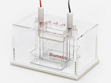
Modern PAGE systems—such as the Biometra Eco-Line and the Biometra Minigel-Twin from Analytik Jena—offer users significant advantages in terms of ease of use, flexibility, and efficiency. The Biometra Eco-Line, with its modular tank system, allows for easy adaptation to different applications and gel sizes, and includes an optional cooling function for reliable separation of sensitive samples. In contrast, the Biometra Minigel-Twin stands out with its compact design, simple handling, and particularly resource-efficient operation, requiring only minimal buffer volumes.

The newsletter of Analytik Jena frequently keeps you posted about:
- News
- Trends and developments
- Events